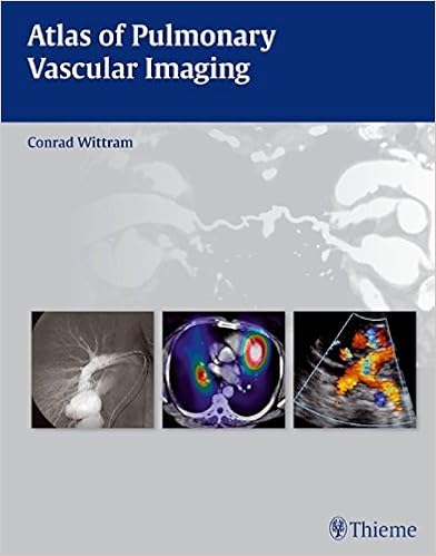
By Conrad Wittram
Packed with distinct, basically categorised radiologic photographs on each web page,
this lavishly illustrated atlas teaches readers the best way to establish and speedy
diagnose the spectrum of pulmonary vascular pathologies utilizing the whole variety of
imaging modalities. each one concise but entire bankruptcy presents systematic
coverage of the imaging manifestations of universal, unusual, and infrequent ailments.
Explanatory textual content vitamins every one top quality snapshot to focus on basically the main
relevant, must-know information.
Features:
- In-depth assurance of ways the pulmonary vessels are
plagued by congenital anomalies, cardiac disorder, emboli, in situ thrombosis,
vasculitis, tumors, aneurysms, and different key lung vessel pathologies - 359 high-resolution radiologic pictures reveal a
wide array of imaging modalities, from radiography, angiography, and
multislice CT, to MRI, ultrasound, and nuclear imaging - Succinct bullet-point structure permits fast and simple
reference - High-quality angiogiographic and correlative CT
photographs and instructive drawings illustrate the diagnostic standards of
pulmonary embolism - Tips on how you can realize pulmonary embolism mimics,
such as partial quantity and flow-related artifacts
This image-rich quantity is
ideal for clinicians, fellows, and citizens in radiology, breathing drugs,
emergency drugs, cardiology, and cardiothoracic surgical procedure as either an academic instrument and a ordinary reference for daily practice.
Read Online or Download Atlas of pulmonary vascular imaging PDF
Similar pulmonary & thoracic medicine books
Endothelium : molecular aspects of metabolic disorders
The functionality and lifestyles span of endothelial cells have a wide influence upon the standard and expectancy of an individual's existence. in the course of low perfusion, the variation of other cells to hypoxia precipitate the competitive development of ailments. even though the medical reports have convincingly proven that endothelial disorder happens at any time when the organic features or bioavailability of nitric oxide are impaired, in some of these eventualities, the position of endothelial cell-destructive approach cross-talk is but poorly understood.
This ebook offers a concise synthesis of the present wisdom and up to date advances within the constitution, association and useful function of the cytoskeleton in endothelial cells. specific awareness has been given to the several good points of the rules of vascular functionality mediated by means of the endothelium.
Now in an absolutely revised and up to date 6th version, Dr. Light's vintage textual content, Pleural illnesses, supplies much more concentrated content material at the pathophysiology, scientific manifestations, analysis, and administration of pleural illnesses. The text’s ordinary, single-author standpoint combines procedural services, insights on fresh technical advances, and transparent concepts for either analysis and therapy.

Nutritional Management of Inflammatory Bowel Diseases: A Comprehensive Guide
This publication is a state-of-the artwork evaluation for clinicians and dieticians with an curiosity in food and inflammatory bowel ailments (Crohn’s illness, ulcerative colitis). the quantity covers new info approximately nutritional danger elements for Crohn’s disorder and ulcerative colitis, examines the organization among vitamin and microbiome, describes a few of the diets within the administration of those illnesses, and discusses macro- and micronutrient deficiency that happens in such sufferers.
Extra info for Atlas of pulmonary vascular imaging
Sample text
6 (A) Illustration of a normal secondary pulmonary lobule and bronchovascular bundle. The secondary pulmonary lobule is represented by the hexagon; in the center are the bronchiole (green ring) and arteriole (red circle); in the periphery are pulmonary venules (short, fat arrow). To the right are cross-sections of a bronchus (arrowhead) and pulmonary artery (arrow). (B) Illustration of interstitial pulmonary edema. There is an increase in the attenuation of the lung parenchyma, but the vessels are still easily seen; this is ground-glass opacification.
Fig. 18 Normal selective digital bronchial artery angiogram in a 27-year-old man. The right intercostobronchial artery branches to the right bronchus and extends superiorly to the right ribs. 1 Anatomy origins include the aortic arch, internal mammary artery, thyrocervical trunk, subclavian artery, costocervical trunk, brachiocephalic artery, pericardiacophrenic artery, inferior phrenic artery, and abdominal aorta. 5 mm in diameter at their origin. 5 mm is considered abnormal. Suggested Reading Cory RA, Valentine EJ.
Fig. 8 (A–C) Arteriovenous malformation in an 83-year-old woman. (A) Chest radiograph demonstrates a serpinginous opacity overlying the left upper lobe (arrow). 2 Congenital Anomalies Fig. 8 (Continued) Arteriovenous malformation in an 83-year-old woman. (B) MIP CT demonstrates a peripheral abnormality consistent with an arteriovenous malformation (arrow). (C) Curved reformatted CT image demonstrates that the abnormality (arrow) is fed directly from a branch of the pulmonary artery (PA) and drains directly into the left atrium (LA), proving that it is an arteriovenous malformation.